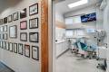
Clinic
Ladies and gentlemen, at your disposal we put a dental center with offices equipped with state-of-the-art equipment, making it possible to perform an all manner of dental procedures in a safe and painless manner.
Our own X-ray laboratory allows us to take a wide range of necessary radiological images.


Equipment

The OMS dental units can be distinguished by solutions that enable the dental team to take advantage of all the principles of ergonomics when performing dental procedures.

GENDEX, a system of digital RadioVisioGraphy, makes it possible to take radiological images, X-rays in digital quality, with minimal dose of radiation exposure, safe for the patient.

GENDEX, an intra-oral camera, facilitates not only the diagnosis making, but also the planning of proper treatment.

Surgical microscopes increase the precision and allow us to cure teeth that were earlier earmarked for extraction due to their complicated build of root canal system, or due to unsuccessful primary treatment (broken instrument in the root canal, or perforation).

VDW Gold endodontic micromotor, with a built-in device for measuring the length of root canals, makes it possible to significantly shorten the duration of the whole procedure, and has an influence on the successful outcome of endodontic treatment.

The system used for filling up the root canals with a thermoplastic, liquid gutta-percha provides an impermeable filling for teeth treated endodontically.

SURTRON, an electrosurgical instrument for performing precise incisions with almost no bleeding, which is often used in the plastic surgery of the frenulum, the gums, or in cases of removing the operculum over erupting teeth.

Dental implant micro motors are used for performing procedures in a safe manner.

Autoclave handles the correct instrument sterilization cycle, ensuring the safety of our Patients.

Swiss dental loupes allow us to perform all dental procedures with pinpoint precision.

The Wand, a computer system, ensures safety and comfort of patients when administering anesthesia.

The face bow and Armann Girbach, an articulator, allow for an individual registration of the occlusion and a precise preparation of the prosthetic device.

The cone beam volumetric tomography (CBCT) is currently a widely used element of 3D imaging diagnostics used in dental surgery. It serves as a great supplement to intra-oral X-rays as well as panoramic x-rays.

Thanks to the application of an ultramodern intraoral scanner, we are able to craft beautiful smiles with computer-based precision, without the necessity of taking impressions. Thanks to a special camera, your dentition is scanned and then saved in our computer.

A camera equipped with a macro lighting system makes it possible to take high-resolution intra-oral images which help with the preparation of the treatment plan as well as the contact with patients.